Bursitis Of The Foot Joint Inflammation
Overview
Is bursitis in your heel or ankle causing pain and discomfort that is affecting your ability to participate in sports, exercise or even possibly beginning to take a toll of your work and life in general? Heel bursitis can be extremely painful and debilitating, what more the heel is such a hard area of the body to properly rest as we are constantly on our feet. This may cause extreme emotional stress on even the most strongly minded individual.
Causes
Occasionally the bursal sac can become inflamed and painful. Pain to the region is worse typically with initial weight bearing activity such as rising from bed in the morning. Swelling and warmth to the region are common. Clinical examination shows pain to palpation at the retrocalcaneus at a level just before the Achilles tendon. Increase pressure and friction of the Achilles tendon across the retrocalcaneal region is the cause of this bursitis. A high arch, tight Achilles tendon or bone spur appear to be some of the main causes of this problem. With a high arch the back portion of the calcaneus abnormally projects into the Achilles tendon region.
Symptoms
When the bursa becomes inflamed after an injury, symptoms usually develop suddenly. When the bursa develops without an injury, symptoms may develop gradually. With both posterior and anterior Achilles tendon bursitis, symptoms usually include swelling and warmth at the back of the heel. A minimally red, swollen, tender spot develops on the back of the heel. When the inflamed bursa enlarges, it appears as a red lump under the skin of the heel and causes pain at and above the heel. If posterior Achilles tendon bursitis becomes chronic, the swelling may become hard, fluid-filled, and red or flesh-colored.
Diagnosis
When a patient has pain in a joint, a careful physical examination is needed to determine what type of movement is affected and if there is any swelling present. Bursitis will not show up on x-rays, although sometimes there are also calcium deposits in the joint that can be seen. Inserting a thin needle into the affected bursa and removing (aspirating) some of the synovial fluid for examination can confirm the diagnosis. In most cases, the fluid will not be clear. It can be tested for the presence of microorganisms, which would indicate an infection, and crystals, which could indicate gout. In instances where the diagnosis is difficult, a local anesthetic (a drug that numbs the area) is injected into the painful spot. If the discomfort stops temporarily, then bursitis is probably the correct diagnosis.
Non Surgical Treatment
Medications may be used to reduce the inflammation and pain of retrocalcaneal bursitis. Nonsteroidal anti-inflammatory drugs such as ibuprofen, naproxen and ketoprofen can be purchased without a prescription and used to treat mild to moderate pain. These drugs are often used in combination with a physical therapy program or other retrocalcaneal bursitis treatments.
Surgical Treatment
Bursectomy is a surgical procedure used to remove an inflamed or infected bursa, which is a fluid-filled sac that reduces friction between tissues of the body. Because retrocalcaneal bursitis can cause chronic inflammation, pain and discomfort, bursectomy may be used as a treatment for the condition when it is persistent and cannot be relived with other treatments. During this procedure, a surgeon makes small incisions so that a camera may be inserted into the joint. This camera is called an arthroscope. Another small incision is made so that surgical instruments can be inserted to remove the inflamed bursa.
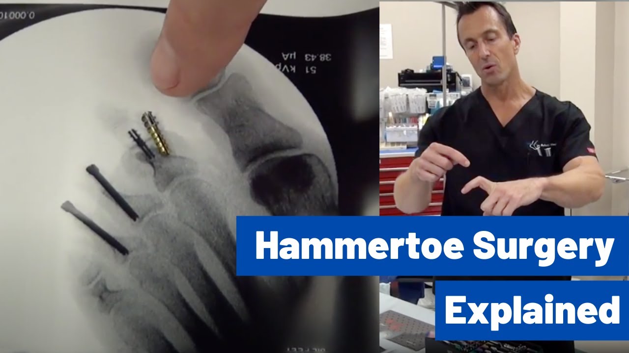
Hammer Toe Surgery Foot Problems
 Overview
The hammertoe condition is usually irreversible, but often its progression can be slowed or halted. You should visit a Podiatrist if the toe becomes painful and you have difficulty walking. A Podiatrist will be able to provide advice and treatment including padding the bony top-part of your hammertoe to relieve pain or to tape your toes as a way to change their position. Podiatrists have an important role to play in preventing and managing foot problems. Prompt action is important. Problems which are left without assessment or treatment may result in major health risks.
Causes
Hammertoe and mallet toe have been linked to certain shoes. High-heeled shoes or footwear that's too tight in the toe box can crowd your toes into a space that's not large enough for them to lie flat. This curled toe position may eventually persist even when you're barefoot. Trauma. An injury in which you stub, jam or break a toe may make it more likely for that digit to develop hammertoe or mallet toe. Nerve injuries or disorders. Hammertoe and mallet toe are more common in people who have nerve damage in their feet, which often occurs with such medical problems as a stroke or diabetes.
 Symptoms
Well-developed hammertoes are distinctive due to the abnormal bent shape of the toe. However, there are many other common symptoms. Some symptoms may be present before the toe becomes overly bent or fixed in the contracted position. Often, before the toe becomes permanently contracted, there will be pain or irritation over the top of the toe, particularly over the joint. The symptoms are pronounced while wearing shoes due to the top of the toe rubbing against the upper portion of the shoe. Often, there is a significant amount of friction between the toe and the shoe or between the toe and the toes on either side of it. The corns may be soft or hard, depending on their location and age. The affected toe may also appear red with irritated skin. In more severe cases, blisters or open sores may form. Those with diabetes should take extra care if they develop any of these symptoms, as they could lead to further complications.
Diagnosis
Your healthcare provider will examine your foot, checking for redness, swelling, corns, and calluses. Your provider will also measure the flexibility of your toes and test how much feeling you have in your toes. You may have blood tests to check for arthritis, diabetes, and infection.
Non Surgical Treatment
You should seek medical advice if you have a hammer toe. Here are some things you can do in the meantime. None of these things will cure the hammer toe, but they may relieve the pain and discomfort. Only wear shoes that are high and broad across the toes. There should be at least 1.5 cm of space between your longest toe and the tip of the shoe. Keep in mind that this could be either your big toe or your second toe. Don't wear heels higher than 5 cm. Wear the appropriate shoe for the activity you are doing. You can buy non-medicated hammer toe pads. They fit around the pointy top of the toe joint and help relieve painful pressure. Gently massaging the toe may help relieve pain. Put ice packs wrapped in cloth on the hammer toe to reduce painful swelling.
Surgical Treatment
For the surgical correction of a rigid hammertoe, the surgical procedure consists of removing Hammer toes the damaged skin where the corn is located. Then a small section of bone is removed at the level of the rigid joint. The sutures remain in place for approximately ten days. During this period of time it is important to keep the area dry. Most surgeons prefer to leave the bandage in place until the patient's follow-up visit, so there is no need for the patient to change the bandages at home. The patient is returned to a stiff-soled walking shoe in about two weeks. It is important to try and stay off the foot as much as possible during this time. Excessive swelling of the toe is the most common patient complaint. In severe cases of hammertoe deformity a pin may be required to hold the toe in place and the surgeon may elect to fuse the bones in the toe. This requires several weeks of recovery.
Over-Pronation Of The Feet Causes
Overview
You know when we walk in the sand on the beach? Well, that?s how nature meant us to walk! You will notice how the sand forms a complete support under the foot. Unfortunately, instead of soft natural surfaces, we walk mainly on hard surfaces like pavements and floors. These hard surfaces cause the foot to roll inwards and the arches to flatten to gain ground contact. This condition is called ?over-pronation? and it affects an estimated 70% of the population. Over-pronation can lead to various foot problems such as heel pain and ball of foot pain. Because our feet are the foundation of our body, poorly aligned feet may also cause problems in other parts of the body, resulting in shin pain, aching legs, knee pain, even lower back pain.
Causes
A common cause of pronation is heredity - we can inherit this biomechanical defect. The second most common cause is due to the way our feet were positioned in the uterus while we were developing; this is called a congenital defect. In either instance, the following occurs in our feet during our development.
Symptoms
Over-pronation is a condition where the arch flattens out which makes the feet roll inward while walking. This condition is also known as flat feet. It imposes extreme additional stresses on the plantar fascia, a fibrous band of tissue which connects the heel to the forefoot. Over-pronation makes walking a painful experience because of the additional strain on the calves, heel and/or back. Treatment for over-pronation involves the use of specially-made orthotics which offers arch support and medial rear foot posting as corrective measures.
Diagnosis
A quick way to see if you over-pronate is to look for these signs. While standing straight with bare feet on the floor, look so see if the inside of your arch or sole touches the floor. Take a look at your hiking or running shoes; look for wear on the inside of the sole. Wet your feet and walk on a surface that will show the foot mark. If you have a neutral foot you should see your heel connected to the ball of your foot by a mark roughly half of width of your sole. If you over-pronate you will see greater than half and up to the full width of your sole.
Non Surgical Treatment
Overpronation is usually corrected with orthotics and/or strengthening exercises for the tibialis posterior. Massage treatment can relieve myofascial trigger points in the tibialis posterior, and other muscles, and address any resulting neuromuscular dysfunction in the leg or foot. Biomechanical correction of overpronation might require orthotics, neuromuscular reeducation, or gait retraining methods, as well. Stretching the gastrocnemius and soleus muscles will reduce hypertonicity in these muscles and also is essential for effective treatment. Because of impacts throughout the remainder of the body, the detrimental effects of overpronation should not be overlooked.
Prevention
Duck stance: Stand with your heels together and feet turned out. Tighten the buttock muscles, slightly tilt your pelvis forwards and try to rotate your legs outwards. You should feel your arches rising while you do this exercise.
Calf stretch:Stand facing a wall and place hands on it for support. Lean forwards until stretch is felt in the calves. Hold for 30 seconds. Bend at knees and hold for a further 30 seconds. Repeat 5 times.
Golf ball:While drawing your toes upwards towards your shins, roll a golf ball under the foot between 30 and 60 seconds. If you find a painful point, keep rolling the ball on that spot for 10 seconds.
Big toe push: Stand with your ankles in a neutral position (without rolling the foot inwards). Push down with your big toe but do not let the ankle roll inwards or the arch collapse. Hold for 5 seconds. Repeat 10 times. Build up to longer times and fewer repetitions.
Ankle strengthener: Place a ball between your foot and a wall. Sitting down and keeping your toes pointed upwards, press the outside of the foot against the ball, as though pushing it into the wall. Hold for 5 seconds and repeat 10 times.
Arch strengthener: Stand on one foot on the floor. The movements needed to remain balanced will strengthen the arch. When you are able to balance for 30 seconds, start doing this exercise using a wobble board.
Living With Calcaneal Apophysitis
Overview
Severs disease (calcaneal apophysitis) is a self-limiting condition seen in physically active children. Although there is controversy about the radiographic appearance, some reports propose the importance of fragmentation of the secondary nucleus in the diagnosis of Severs disease. We studied secondary nucleus of the calcaneus with ultrasonography. Twenty-one symptomatic heels of 14 children were examined. All these heels showed fragmentation of the secondary nucleus on both conventional radiograph and sonography. Ultrasonographic examination also showed 2 retrocalcaneal bursitis. Our initial data showed that sonography may be valuable in the diagnosis of Severs disease.
Causes
Heel pain is very common in children because of the very nature of their growing feet and legs. In children, the heel bone (the calcaneus) is not fully developed until the age of 14 or older. Until then, new bone is forming at the growth plate of the foot (the apophysis, located at the back of the heel), an area which is softer than others due to its role in accommodating the growth. Repetitive stress on the growth plate due to walking, running and sports causes inflammation in the heel area. Because the heel's growth plate is sensitive, repeated running and pounding on hard surfaces can result in pediatric heel pain. Children and adolescents involved in football, soccer, running or basketball are especially vulnerable. Over-pronation (fallen arches and rolling inwards of the feet) will increase the stress on the growth plate and is therefore a significant cause and a major contributing factor to heel pain in children.
Symptoms
The most prominent symptom of Sever's disease is heel pain which is usually aggravated by physical activity such as walking, running or jumping. The pain is localised to the posterior and plantar side of the heel over the calcaneal apophysis. Sometimes, the pain may be so severe that it may cause limping and interfere with physical performance in sports. External appearance of the heel is almost always normal, and signs of local disease such as edema, erythema (redness) are absent. The main diagnostic tool is pain on medial- lateral compression of the calcaneus in the area of growth plate, so called squeeze test. Foot radiographs are usually normal. Therefore the diagnosis of Sever's disease is primarily clinical.
Diagnosis
Your Podiatrist or Physiotherapist will assist in diagnosing the injury and the extent of the damage. From this, they will develop a management plan which may include rest or activity modification, soft tissue treatment such as massage and stretching, correction of biomechanics through heel raises or orthoses and the progression through a series of specific strengthening exercises.
Non Surgical Treatment
The treatment of Sever's disease should be individualized. The most important first steps in the treatment of Sever's disease are activity modification (including rest and sometimes crutches) and good shoes. Further treatment may include icing to decrease pain around the calcaneal apophysis, stretching and strengthening exercises, shoe orthotics or medications to relieve pain. Rarely, a removable cast is necessary to completely rest the foot.
Achilles Tendon Rupture Pain Level
Overview
 The Achilles tendon affects your ability to do everything from walking to playing competitive sports. When a patient overstretches his or her Achilles tendon, it can result in a full or partial tear in the tendon, also known as a rupture. In addition to causing a great deal of pain, ruptures can have a profoundly negative impact on your quality of life and prevent you from performing activities you once enjoyed. Because these injuries tend to worsen with time, it?s important to contact a board certified orthopedic surgeon for immediate attention after an Achilles tendon tear.
Causes
Often an Achilles rupture can occur spontaneously without any prodromal symptoms. Unfortunately the first "pop" or "snap" that you experience is your Achilles tendon rupture. Achilles tendon rupture most commonly occurs in the middle-aged male athlete (the weekend warrior who is engaging in a pickup game of basketball, for example). Injury often occurs during recreational sports that require bursts of jumping, pivoting, and running. Most often these are tennis, racquetball, squash, basketball, soccer, softball and badminton. Achilles rupture can happen in the following situations. You make a forceful push-off with your foot while your knee is straightened by the powerful thigh muscles. One example might be starting a foot race or jumping. You suddenly trip or stumble, and your foot is thrust in front to break a fall, forcefully over stretching the tendon. You fall from a significant height. It does appear that previous history of Achilles tendonitis results in a degenerative tendon, which can grow weak and thin with age and lack of use. Then it becomes prone to injury or rupture. Certain illnesses (such as arthritis and diabetes) and medications (such as corticosteroids and some antibiotics) can also increase the risk of rupture.
Symptoms
If your Achilles tendon is ruptured you will experience severe pain in the back of your leg, swelling, stiffness, and difficulty to stand on tiptoe and push the leg when walking. A popping or snapping sound is heard when the injury occurs. You may also feel a gap or depression in the tendon, just above heel bone.
Diagnosis
Laboratory studies usually are not necessary in evaluating and diagnosing an Achilles tendon rupture or injury, although evaluation may help to rule out some of the other possibilities in the differential diagnosis. Plain radiography. Radiographs are more useful for ruling out other injuries than for ruling in Achilles tendon ruptures. Ultrasonography of the leg and thigh can help to evaluate the possibility of deep venous thrombosis and also can be used to rule out a Baker cyst, in experienced hands, ultrasonography can identify a ruptured Achilles tendon or the signs of tendinosis. Magnetic resonance imaging (MRI). MRI can facilitate definitive diagnosis of a disrupted tendon and can be used to distinguish between paratenonitis, tendinosis, and bursitis.
Non Surgical Treatment
The other option is to allow your tendon to heal without surgery. In this case, you also need to wear a cast, splint, walking boot, or brace for 6-8 weeks. You also may have different exercises to do. If you are less active or have a chronic illness that prevents surgery, this option may be better for you. 
Surgical Treatment
Some surgeons feel an early surgical repair of the tendon is beneficial. The surgical option was long thought to offer a significantly smaller risk of re-rupture compared to traditional non-operative management (5% vs 15%). Of course, surgery imposes higher relative risks of perioperative mortality and morbidity e.g. infection including MRSA, bleeding, deep vein thrombosis, lingering anesthesia effects, etc.
Posterior Tibial Tendon Dysfunction Exercises
Overview
The posterior tibial tendon serves as one of the major supporting structures of the foot, helping it to function while walking. Posterior tibial tendon dysfunction (PTTD) is a condition caused by changes in the tendon, impairing its ability to support the arch. This results in flattening of the foot. PTTD is often called ?adult acquired flatfoot? because it is the most common type of flatfoot developed during adulthood. Although this condition typically occurs in only one foot, some people may develop it in both feet. PTTD is usually progressive, which means it will keep getting worse, especially if it isn?t treated early. 
Causes
As discussed above, many different problems can create a painful flatfoot. Damage to the posterior tibial tendon is the most common cause of AAFD. The posterior tibial tendon is one of the most important tendons of the leg. It starts at a muscle in the calf, travels down the inside of the lower leg and attaches to the bones on the inside of the foot. The main function of this tendon is to support the arch of your foot when you walk. If the tendon becomes inflamed or torn, the arch will slowly collapse. Women and people over 40 are more likely to develop problems with the posterior tibial tendon. Other risk factors include obesity, diabetes, and hypertension. Having flat feet since childhood increases the risk of developing a tear in the posterior tibial tendon. In addition, people who are involved in high impact sports, such as basketball, tennis, or soccer, may have tears of the tendon from repetitive use. Inflammatory arthritis, such as rheumatoid arthritis, can cause a painful flatfoot. This type of arthritis attacks not only the cartilage in the joints, but also the ligaments that support the foot. Inflammatory arthritis not only causes pain, but also causes the foot to change shape and become flat. The arthritis can affect the back of the foot or the middle of foot, both of which can result in a fallen arch. An injury to the tendons or ligaments in the foot can cause the joints to fall out of alignment. The ligaments support the bones and prevent them from moving. If the ligaments are torn, the foot will become flat and painful. This more commonly occurs in the middle of the foot (Lisfranc injury), but can also occur in the back of the foot. Injuries to tendons of the foot can occur either in one instance (traumatically) or with repeated use over time (overuse injury). Regardless of the cause, if tendon function is altered, the forces that are transmitted across joints in the foot are changed and this can lead to increased stress on joint cartilage and ligaments. In addition to tendon and ligament injuries, fractures and dislocations of the bones in the midfoot can also lead to a flatfoot deformity. People with diabetes or with nerve problems that limits normal feeling in the feet, can have collapse of the arch or of the entire foot. This type of arch collapse is typically more severe than that seen in patients with normal feeling in their feet. In addition to the ligaments not holding the bones in place, the bones themselves can sometimes fracture and disintegrate without the patient feeling any pain. This may result in a severely deformed foot that is very challenging to correct with surgery. Special shoes or braces are the best method for dealing with this problem.
Symptoms
Often, this condition is only present in one foot, but it can affect both. Adult acquired flatfoot symptoms vary, but can swelling of the foot's inner side and aching heel and arch pain. Some patients experience no pain, but others may experience severe pain. Symptoms may increase during long periods of standing, resulting in fatigue. Symptoms may change over time as the condition worsens. The pain may move to the foot's outer side, and some patients may develop arthritis in the ankle and foot.
Diagnosis
Clinicians need to recognize the early stage of this syndrome which includes pain, swelling, tendonitis and disability. The musculoskeletal portion of the clinical exam can help determine the stage of the disease. It is important to palpate the posterior tibial tendon and test its muscle strength. This is tested by asking patient to plantarflex and invert the foot. Joint range of motion is should be assessed as well. Stiffness of the joints may indicate longstanding disease causing a rigid deformity. A weightbearing examination should be performed as well. A complete absence of the medial longitudinal arch is often seen. In later stages the head of the talus bone projects outward to the point of a large "lump" in the arch. Observing the patient's feet from behind shows a significant valgus rotation of the heel. From behind, the "too many toes" sign may be seen as well. This is when there is abducution of the forefoot in the transverse plane allowing the toes to be seen from behind. Dysfunction of the posterior tibial tendon can be assessed by asking the patient to stand on his/her toes on the affected foot. If they are unable to, this indicates the disease is in a more advanced stage with the tendon possibly completely ruptured.
Non surgical Treatment
Orthoses (insoles, functional orthoses, ankle supports, braces, ankle foot orthoses (AFOs)) - are usually custom-made to increase the functional stability of the foot and improve the mechanical properties of the tendon as well as reducing the actual degree of strain on the tendon. This reduces pain and inflammation. Physiotherapy - exercises and physiotherapy are often used to increase mobility, strengthen the tendon itself, stretch your Achilles tendon as well as reduce pain. Once the tendon has been stretched (stage one), the heel starts rolling outwards. Total immobilisation in a cast may help the symptoms to subside and prevent progression of the deformity in a smaller percentage of patients. Long-term use of orthoses may help stop progression of the deformity and reduce pain without surgery. Non-surgical treatment is unlikely to prevent progression to stage three and four but may be chosen by some patients who either are unsuitable for surgery or prefer not to have surgery. 
Surgical Treatment
Surgery should only be done if the pain does not get better after a few months of conservative treatment. The type of surgery depends on the stage of the PTTD disease. It it also dictated by where tendonitis is located and how much the tendon is damaged. Surgical reconstruction can be extremely complex. Some of the common surgeries include. Tenosynovectomy, removing the inflamed tendon sheath around the PTT. Tendon Transfer, to augment the function of the diseased posterior tibial tendon with a neighbouring tendon. Calcaneo-osteotomy, sometimes the heel bone needs to be corrected to get a better heel bone alignment. Fusion of the Joints, if osteoarthritis of the foot has set in, fusion of the joints may be necessary.